Copyright
©The Author(s) 2015.
World J Cardiol. Dec 26, 2015; 7(12): 861-874
Published online Dec 26, 2015. doi: 10.4330/wjc.v7.i12.861
Published online Dec 26, 2015. doi: 10.4330/wjc.v7.i12.861
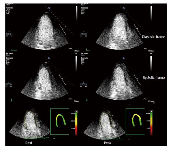
Figure 6 Myocardial contrast stress echocardiography.
Apical three-chamber view of a patient with previous by-pass graft (left internal mammary artery graft onto left anterior descending coronary artery). Baseline diastolic and systolic frames on the left, top and intermediate rows, respectively; peak stress diastolic and systolic frames on the right, top and intermediate rows, respectively. Baseline and peak stress myocardial perfusion parametric quantification are displayed, respectively, in the bottom left and right panels. At baseline, akinesis of the infero-apical region is evident, which is concordant with a transmural defect of perfusion of the same region. At peak stress, no new wall motion abnormalities are detected, whereas parametric quantification of myocardial perfusion shows a large transmural defect of perfusion of all the apical regions and a subendocardial defect of perfusion of middle and basal anterior septum. A critical stenosis of distal mammary graft anastomosis was found on coronary angiography.
- Citation: Barletta G, Del Bene MR. Myocardial perfusion echocardiography and coronary microvascular dysfunction. World J Cardiol 2015; 7(12): 861-874
- URL: https://www.wjgnet.com/1949-8462/full/v7/i12/861.htm
- DOI: https://dx.doi.org/10.4330/wjc.v7.i12.861


