Copyright
©The Author(s) 2015.
World J Cardiol. Nov 26, 2015; 7(11): 776-783
Published online Nov 26, 2015. doi: 10.4330/wjc.v7.i11.776
Published online Nov 26, 2015. doi: 10.4330/wjc.v7.i11.776
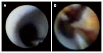
Figure 1 Coronary angioscopy images of ordinary neoin-tima and neoatherosclerosis.
A: Coronary angioscopy reveals ordinary neointima as a white and smooth membranous structure; B: The neointima appears as atheromatous and yellow, occasionally disrupted with thrombus formation.
- Citation: Komiyama H, Takano M, Hata N, Seino Y, Shimizu W, Mizuno K. Neoatherosclerosis: Coronary stents seal atherosclerotic lesions but result in making a new problem of atherosclerosis. World J Cardiol 2015; 7(11): 776-783
- URL: https://www.wjgnet.com/1949-8462/full/v7/i11/776.htm
- DOI: https://dx.doi.org/10.4330/wjc.v7.i11.776


