Copyright
©The Author(s) 2015.
World J Cardiol. Oct 26, 2015; 7(10): 700-702
Published online Oct 26, 2015. doi: 10.4330/wjc.v7.i10.700
Published online Oct 26, 2015. doi: 10.4330/wjc.v7.i10.700
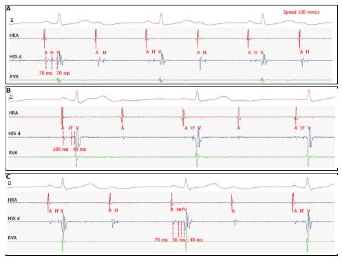
Figure 1 Surface electrocardiograms with intra-cardiac electrocardiograms in sweep speed of 100 mm/s.
Surface ECG shows 2:1 atrioventricular block with a right bundle branch block. A: Intra-cardiac ECG was located the right ventricular apex (RVA), His bundle (HIS), and right ventricular apex (RVA). Surface ECG revealed 2:1 atrio-ventricular block. Intra-cardiac ECG revealed infra-hisian block and short A-H interval (70 ms); B: Intra-cardiac ECG revealed supra-hisian block and long A-H interval (100 ms); C: The intracardiac ECG shows the splitting of the His potential (H-H’) during AV conduction and only the proximal activation of His potential during the AV block, which means an intra-hisian block. A: Potential of right atrium; H: Potential of proximal His bundle; H’: Potential of distal His bundle; V: Potential of right ventricle; ECG: Electrocardiogram.
- Citation: Hong SP, Park YW, Lee YS. Intra-His bundle block in 2:1 atrioventricular block. World J Cardiol 2015; 7(10): 700-702
- URL: https://www.wjgnet.com/1949-8462/full/v7/i10/700.htm
- DOI: https://dx.doi.org/10.4330/wjc.v7.i10.700


