Copyright
©The Author(s) 2015.
World J Cardiol. Oct 26, 2015; 7(10): 583-593
Published online Oct 26, 2015. doi: 10.4330/wjc.v7.i10.583
Published online Oct 26, 2015. doi: 10.4330/wjc.v7.i10.583
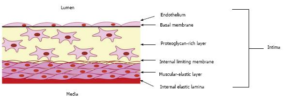
Figure 1 Schema showing the organization of the intima of the arterial wall.
The proteoglycan-rich layer which contains a heterogeneous population of cells, including macrovascular pericytes, is located just below the endothelial monolayer. Intimal pericytes form a network of cells interconnected through gap junctions. The muscular-elastic layer, formed by elongated contractile smooth muscular cells, lies below the proteoglycan-rich layer.
- Citation: Ivanova EA, Bobryshev YV, Orekhov AN. Intimal pericytes as the second line of immune defence in atherosclerosis. World J Cardiol 2015; 7(10): 583-593
- URL: https://www.wjgnet.com/1949-8462/full/v7/i10/583.htm
- DOI: https://dx.doi.org/10.4330/wjc.v7.i10.583


