Copyright
©2014 Baishideng Publishing Group Inc.
World J Cardiol. Sep 26, 2014; 6(9): 968-984
Published online Sep 26, 2014. doi: 10.4330/wjc.v6.i9.968
Published online Sep 26, 2014. doi: 10.4330/wjc.v6.i9.968
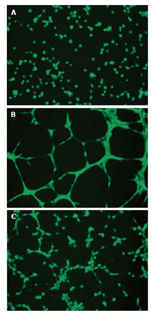
Figure 7 Conditioned medium collected from human ARPE-19 cells exposed to Ang II promotes tube formation in choroidal microvascular endothelial through AT1 activation[140].
Cells were exposed to: (1) Ang II alone; or (2) Ang II in combination with candesartan for 24 h, supernatants were collected after treatment and human choroidal microvascular endothelial (cECs) were treated with the supernatants for 24 h. Thereafter, cells were trypsinized and then seeded (42000 cells/cm2) on a 24-well polystyrene plate coated with Geltrex™ (50 μL/cm2) according to the manufacturer’s protocol followed by incubation in EBM medium for 24 h at 37 °C in 5% CO2. At 16 h post-seeding, 2 μg/mL of Calcein, AM (Invitrogen, Cat # C3099), was added directly to the culture well and allowed to incubate for 20 min (37 °C, 5% CO2). Cells were visualized using a fluorescence microscope. A: Control; B: cECs exposed to conditioned medium from Ang II-treated ARPE-19 cells; C: cECs treated with medium collected from treated retinal pigment epithelium cells. EBM: Endothelial cell basal; AM: Acetoxymethyl.
- Citation: Marin Garcia PJ, Marin-Castaño ME. Angiotensin II-related hypertension and eye diseases. World J Cardiol 2014; 6(9): 968-984
- URL: https://www.wjgnet.com/1949-8462/full/v6/i9/968.htm
- DOI: https://dx.doi.org/10.4330/wjc.v6.i9.968


