Copyright
©2014 Baishideng Publishing Group Inc.
World J Cardiol. Sep 26, 2014; 6(9): 968-984
Published online Sep 26, 2014. doi: 10.4330/wjc.v6.i9.968
Published online Sep 26, 2014. doi: 10.4330/wjc.v6.i9.968
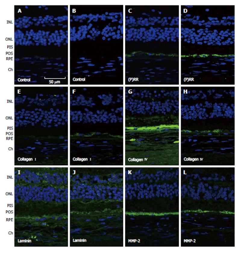
Figure 4 Representative immunofluorescent double staining of prorenin receptor, collagen types I and IV, laminin and matrix metalloproteinase-2 (green) and nuclei (bleu) in retina sections from human donor eyes with no known eye disease (B, D, F, H, J and L), and human donor eyes with dry age-related macular degeneration and hypertension (A, C, E, G, I and K)[121].
Negative controls were generated by omission of the primary antibody (A and B). Sections were analyzed by using confocal microscopy (original magnification, × 40). INL: Inner sections were analyzed with a confocal microscope at a magnification of × 40. INL: Inner nuclear layer; ONL: Outer nuclear layer; MMP: Matrix metalloproteinase; PIS: Photoreceptor inner segments; POS: Photoreceptor outer segments; RPE: Retinal pigment epithelium; Ch: Choroid.
- Citation: Marin Garcia PJ, Marin-Castaño ME. Angiotensin II-related hypertension and eye diseases. World J Cardiol 2014; 6(9): 968-984
- URL: https://www.wjgnet.com/1949-8462/full/v6/i9/968.htm
- DOI: https://dx.doi.org/10.4330/wjc.v6.i9.968


