Copyright
©2014 Baishideng Publishing Group Inc.
World J Cardiol. Aug 26, 2014; 6(8): 836-846
Published online Aug 26, 2014. doi: 10.4330/wjc.v6.i8.836
Published online Aug 26, 2014. doi: 10.4330/wjc.v6.i8.836
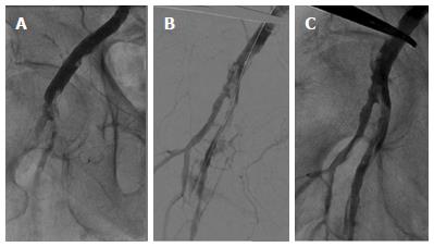
Figure 5 Post-transcatheter aortic valve implantation arterial dissection.
Post- transcatheter aortic valve implantation procedure, angiography of the right iliac-femoral axis via the contralateral groin showing a dissection of the right common femoral artery extending proximally to the external iliac artery and determining distally an occlusion of the superficial femoral artery (A); digital subtraction angiography after reaching true lumen by a .035” wire by the retrograde approach and peripheral balloon dilation (6.0 mm x 120 mm Admiral Xtreme, Invatec, Roncadelle, Italy) to 6 atm (B); final angiography after stenting (6.0 mm x 80 mm and 9.0 mm x 60 mm Lifestent Vascular Stent, BARD Peripheral Vascular, AZ, United States) and post-dilation (5.0 mm x 80 mm and 6.0 mm x 120 mm Admiral Xtreme; Invatec, Roncadelle, Italy) showing an optimal antegrade flow in the right iliac-femoral artery (C).
- Citation: Dato I, Burzotta F, Trani C, Crea F, Ussia GP. Percutaneous management of vascular access in transfemoral transcatheter aortic valve implantation. World J Cardiol 2014; 6(8): 836-846
- URL: https://www.wjgnet.com/1949-8462/full/v6/i8/836.htm
- DOI: https://dx.doi.org/10.4330/wjc.v6.i8.836


