Copyright
©2014 Baishideng Publishing Group Inc.
World J Cardiol. Aug 26, 2014; 6(8): 836-846
Published online Aug 26, 2014. doi: 10.4330/wjc.v6.i8.836
Published online Aug 26, 2014. doi: 10.4330/wjc.v6.i8.836
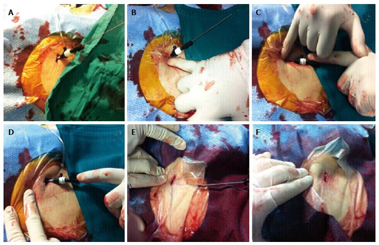
Figure 1 Pre-closure technique for hemostasis in transcatheter aortic valve implantation procedures.
After angiography-guided puncture of the anterior wall of the common femoral artery (CFA) and the insertion of a 6 F sheath, the preparation of vascular access for large sheath insertion (≥ 18 F) consists of the enlargement of the access site by the insertion of a 9 F sheath (A) and dilation of the subcutaneous tissue anteriorly (B) and posteriorly to the sheath (C), using one finger. Such a maneuver should achieve a less traumatic flaring of cutaneous and subcutaneous tissues at the vascular access site and create appropriate space for both large sheath introduction at the beginning of the procedure and optimal fastening of knots over the arterial wall at procedure end (D). After 9 F sheath removal, the suture-mediated vascular closure device is inserted in the correct position, the needles are unlocked and pulled through the arterial wall (E). At the end of transcatheter aortic valve implantation, the sheath and the guide wire are removed, the sutures are fastened individually with a sliding knot and a knot pusher is used to ensure approximation of the knot to the surface of the vessel wall. Vascular suture ends are cut well beneath the surface of the skin and an optimal closure of vascular access is obtained by a single cutaneous suture without residual bleeding (F).
- Citation: Dato I, Burzotta F, Trani C, Crea F, Ussia GP. Percutaneous management of vascular access in transfemoral transcatheter aortic valve implantation. World J Cardiol 2014; 6(8): 836-846
- URL: https://www.wjgnet.com/1949-8462/full/v6/i8/836.htm
- DOI: https://dx.doi.org/10.4330/wjc.v6.i8.836


