Copyright
©2014 Baishideng Publishing Group Inc.
World J Cardiol. Jul 26, 2014; 6(7): 689-691
Published online Jul 26, 2014. doi: 10.4330/wjc.v6.i7.689
Published online Jul 26, 2014. doi: 10.4330/wjc.v6.i7.689
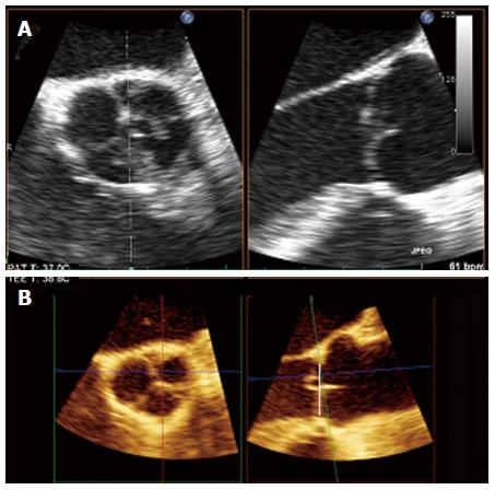
Figure 3 Patient male, 53-year-old.
A: On 2 D there is no clear demonstration of prolapse; B: Multiple plane reconstruction of the aortic valve with 3D transesophageal echocardiography: on reformatted image a prolapse of left coronary cusps is shown (right).
- Citation: Nijs J, Gelsomino S, Kietselaer BB, Parise O, Lucà F, Maessen JG, Meir ML. 3D-echo in preoperative assessment of aortic cusps effective height. World J Cardiol 2014; 6(7): 689-691
- URL: https://www.wjgnet.com/1949-8462/full/v6/i7/689.htm
- DOI: https://dx.doi.org/10.4330/wjc.v6.i7.689


