Copyright
©2014 Baishideng Publishing Group Inc.
World J Cardiol. Jul 26, 2014; 6(7): 653-662
Published online Jul 26, 2014. doi: 10.4330/wjc.v6.i7.653
Published online Jul 26, 2014. doi: 10.4330/wjc.v6.i7.653
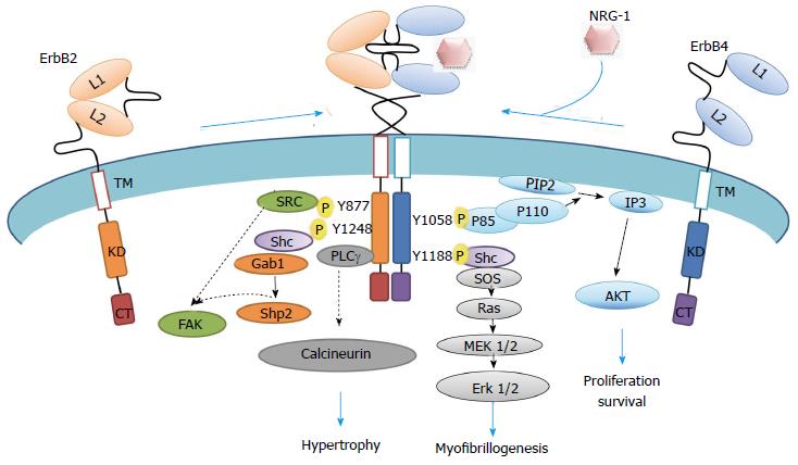
Figure 2 Representation of neuregulin-1-erbB2/erbB4 intracellular signaling cascade.
Schematic representation of active ErbB2/ErbB4 heterodimers through phosphorylation, which phosphosites are docking sites for intracellular molecules involved in pathways that modulate myocyte biology. Specific non-phosphorylated residues interact to PDZ domain proteins. CT: Cytoplasmic tail; KD: Kinase domain; L: Ligand binding site; TM: Transmembrane domain; NRG-1: Neuregulin-1; PIP2: Phosphoinositol-2-phosphate; SOS: Son of sevenless; IP3: Inositol triphosphate; AKT: Thymoma viral oncogene homolog 1, a serine/threonine protein kinase; MEK: Mitogen activated kinase erk kinase; Shc: Src homology domain containing transforming protein; Shp: Protein tyrosine phosphatase; FAK: Focal adhesion kinase; Gab: Binding protein of growth factor bound protein Grb2; P: Phosphorylated tyrosine residues.
- Citation: Vasti C, Hertig CM. Neuregulin-1/erbB activities with focus on the susceptibility of the heart to anthracyclines. World J Cardiol 2014; 6(7): 653-662
- URL: https://www.wjgnet.com/1949-8462/full/v6/i7/653.htm
- DOI: https://dx.doi.org/10.4330/wjc.v6.i7.653


