Copyright
©2014 Baishideng Publishing Group Inc.
World J Cardiol. Jul 26, 2014; 6(7): 585-601
Published online Jul 26, 2014. doi: 10.4330/wjc.v6.i7.585
Published online Jul 26, 2014. doi: 10.4330/wjc.v6.i7.585
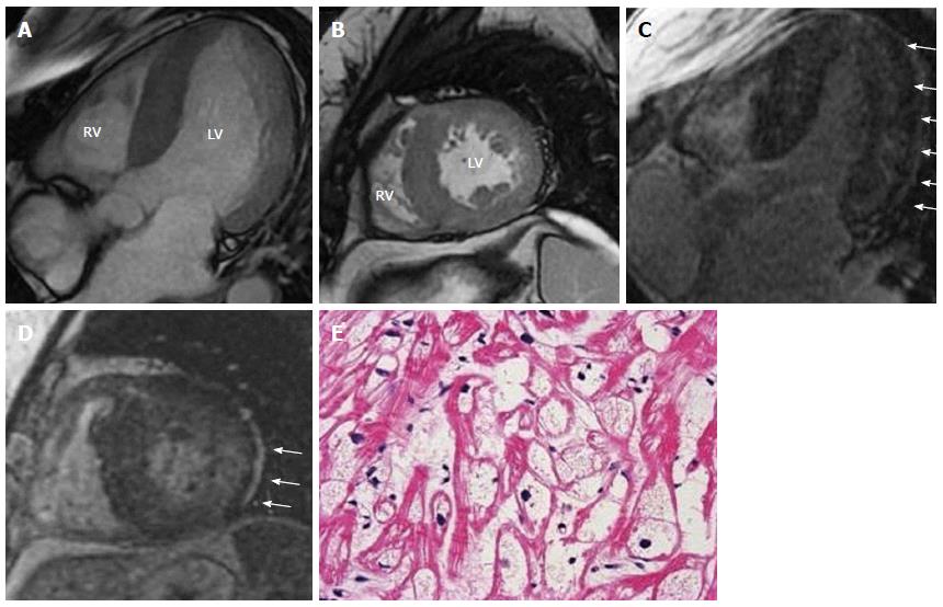
Figure 9 Representative cine- cardiac magnetic resonance (A, B) and late gadolinium enhancement-cardiac magnetic resonance (C, D) images in a 46-year-old female patient with Anderson-fabry disease.
The images show horizontal axis (4-chambers) (A, C) and mid-ventricular short axis (B, D) views. Cine-CMR images reveal diffuse hypertrophy of LV wall. LGE-CMR images show a particular LGE distribution pattern to the infero-lateral mid to basal segments and to mid-myocardial layer (white arrows). E: A sub-endocardial biopsy from RV wall demonstrates interstitial fibrosis and cardiomyocyte hypertrophy with cytoplasmic vacuolization (H-E stain, 40×). LGE: Late gadolinium enhancement; LV: Left ventricular; LGE-CMR: Late gadolinium enhancement-cardiac magnetic resonance; RV: Right ventricular.
- Citation: Satoh H, Sano M, Suwa K, Saitoh T, Nobuhara M, Saotome M, Urushida T, Katoh H, Hayashi H. Distribution of late gadolinium enhancement in various types of cardiomyopathies: Significance in differential diagnosis, clinical features and prognosis. World J Cardiol 2014; 6(7): 585-601
- URL: https://www.wjgnet.com/1949-8462/full/v6/i7/585.htm
- DOI: https://dx.doi.org/10.4330/wjc.v6.i7.585


