Copyright
©2014 Baishideng Publishing Group Inc.
World J Cardiol. Jul 26, 2014; 6(7): 577-584
Published online Jul 26, 2014. doi: 10.4330/wjc.v6.i7.577
Published online Jul 26, 2014. doi: 10.4330/wjc.v6.i7.577
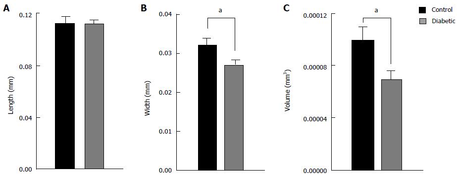
Figure 4 Average dimensions of isolated ventricular myocytes from diabetic and control rat hearts.
Cell length (A) was not different between diabetic (n = 35) and control (n = 19) hearts, whereas cell width (B) and cell volume (C) was reduced. aP < 0.05, diabetic vs control.
- Citation: Ward ML, Crossman DJ. Mechanisms underlying the impaired contractility of diabetic cardiomyopathy. World J Cardiol 2014; 6(7): 577-584
- URL: https://www.wjgnet.com/1949-8462/full/v6/i7/577.htm
- DOI: https://dx.doi.org/10.4330/wjc.v6.i7.577


