Copyright
©2014 Baishideng Publishing Group Inc.
World J Cardiol. Jun 26, 2014; 6(6): 478-494
Published online Jun 26, 2014. doi: 10.4330/wjc.v6.i6.478
Published online Jun 26, 2014. doi: 10.4330/wjc.v6.i6.478
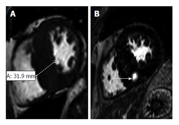
Figure 8 Hypertrophic cardiomyopathy in a 57-year-old man with a 2-year history of exertional dyspnea and chest discomfort who underwent implantable cardioverter-defibrillator placement for primary prevention of sudden cardiac death[105].
A: Short-axis 2D SSFP magnetic resonance imaging (MRI) performed in end diastole shows asymmetric septal hypertrophy with a maximal thickness of 31.9 mm encroaching on the ventricular lumen; B: Short-axis late contrast-enhanced MRI shows a patchy nodular area of enhancement in the hypertrophied septum (arrow) that does not correspond to a coronary artery territory and, therefore, is distinctly different from an infarct scar.
- Citation: Sisakian H. Cardiomyopathies: Evolution of pathogenesis concepts and potential for new therapies. World J Cardiol 2014; 6(6): 478-494
- URL: https://www.wjgnet.com/1949-8462/full/v6/i6/478.htm
- DOI: https://dx.doi.org/10.4330/wjc.v6.i6.478


