Copyright
©2014 Baishideng Publishing Group Inc.
World J Cardiol. Jun 26, 2014; 6(6): 478-494
Published online Jun 26, 2014. doi: 10.4330/wjc.v6.i6.478
Published online Jun 26, 2014. doi: 10.4330/wjc.v6.i6.478
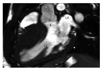
Figure 7 Hypertrophic cardiomyopathy[105].
A: 2D SSFP cardiac magnetic resonance imaging, obtained in end diastole in the long-axis plane of the LV outflow tract (LVOT) in a 17-year-old boy with Hypertrophic cardiomyopathy found at family screening, shows marked asymmetric septal hypertrophy with a ratio of ventricular septal thickness (27 mm, arrow) to inferiolateral wall thickness (9 mm, white arrowhead) of 3:1. Note that the hypertrophied septum encroaches on the LV lumen, causing mild narrowing of the LVOT (black arrowhead). LA: Left atrium; LV: Left ventricle.
- Citation: Sisakian H. Cardiomyopathies: Evolution of pathogenesis concepts and potential for new therapies. World J Cardiol 2014; 6(6): 478-494
- URL: https://www.wjgnet.com/1949-8462/full/v6/i6/478.htm
- DOI: https://dx.doi.org/10.4330/wjc.v6.i6.478


