Copyright
©2014 Baishideng Publishing Group Inc.
World J Cardiol. Jun 26, 2014; 6(6): 478-494
Published online Jun 26, 2014. doi: 10.4330/wjc.v6.i6.478
Published online Jun 26, 2014. doi: 10.4330/wjc.v6.i6.478
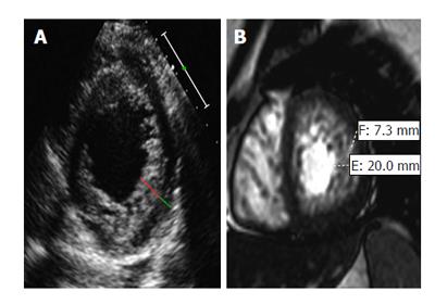
Figure 3 Non-compaction cardiomyopathy in two patients[105].
A: Dilated cardiomyopathy in a 60-year-old man with new-onset congestive heart failure. Short-axis echocardiogram obtained in systole at the level of the left ventricle (LV) shows a two-layered myocardium with a noncompacted (red line) and com¬pacted (green line) layer along the lateral, inferior, and anterior walls and a maximal end-systolic NC: C ratio > 2; B: Symptoms of New York Heart Association Class III heart failure and severely reduced (≤ 35%) LV ejection fraction in a 35-year-old woman. Short-axis 2D SSFP cardiac magnetic resonance imaging obtained in end diastole shows thickening of the noncompacted layer of the LV myocardium, with an NC: C ratio of 2.9. The patient underwent subsequent implantable cardioverter-defibrillator placement for primary prevention of sudden cardiac death.
- Citation: Sisakian H. Cardiomyopathies: Evolution of pathogenesis concepts and potential for new therapies. World J Cardiol 2014; 6(6): 478-494
- URL: https://www.wjgnet.com/1949-8462/full/v6/i6/478.htm
- DOI: https://dx.doi.org/10.4330/wjc.v6.i6.478


