Copyright
©2014 Baishideng Publishing Group Inc.
World J Cardiol. May 26, 2014; 6(5): 345-348
Published online May 26, 2014. doi: 10.4330/wjc.v6.i5.345
Published online May 26, 2014. doi: 10.4330/wjc.v6.i5.345
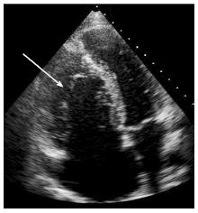
Figure 3 Echocardiography apical 4 chamber view showing the apically displaced tricuspid valve orifice, small right ventricle, large atrialized right ventricle and dilated right atrial.
- Citation: Vette LC, Brugts JJ, McGhie JS, Roos-Hesselink JW. Long-lasting symptoms and diagnostics in a patient with unrecognized right sided heart failure: Why listening to the heart is so important. World J Cardiol 2014; 6(5): 345-348
- URL: https://www.wjgnet.com/1949-8462/full/v6/i5/345.htm
- DOI: https://dx.doi.org/10.4330/wjc.v6.i5.345


