Copyright
©2014 Baishideng Publishing Group Inc.
World J Cardiol. May 26, 2014; 6(5): 314-326
Published online May 26, 2014. doi: 10.4330/wjc.v6.i5.314
Published online May 26, 2014. doi: 10.4330/wjc.v6.i5.314
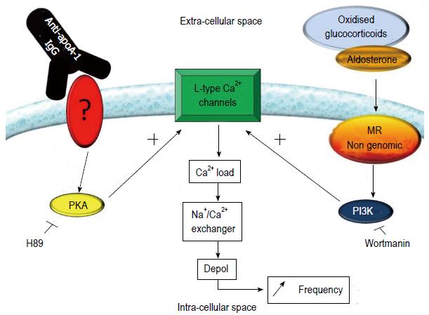
Figure 4 Current understanding of the mechanism by which autoantibodies against apolipoprotein A-1 IgG elicit chronotropic responses in nenonatal rat cardiomyocytes.
Stimulation of the mineralocorticoid receptor (MR), either by aldosterone or oxidized glucocorticoids, induces the downstream activation of PI3K, which in turn activates L-type calcium channels. Anti-apoA-1 IgG has been shown to sensitize the L-type calcium channel in a protein kinase A (PKA)-dependent manner. The PI3K and PKA activated pathways alone are not sufficient to induce an increase in basal contraction rate, when simultaneously activated L-type calcium channels are activated, leading to an increase in intracellular Ca2+. This signal is amplified by the Na+/Ca2+ exchanger, leading to an increase of the prepotential slope of the cells, which ultimately translates into an increased contraction rate.
- Citation: Vuilleumier N, Montecucco F, Hartley O. Autoantibodies to apolipoprotein A-1 as a biomarker of cardiovascular autoimmunity. World J Cardiol 2014; 6(5): 314-326
- URL: https://www.wjgnet.com/1949-8462/full/v6/i5/314.htm
- DOI: https://dx.doi.org/10.4330/wjc.v6.i5.314


