Copyright
©2014 Baishideng Publishing Group Co.
World J Cardiol. Apr 26, 2014; 6(4): 154-174
Published online Apr 26, 2014. doi: 10.4330/wjc.v6.i4.154
Published online Apr 26, 2014. doi: 10.4330/wjc.v6.i4.154
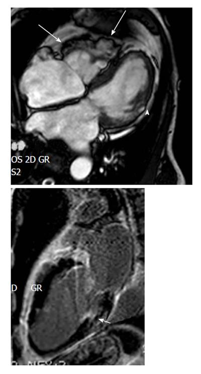
Figure 5 Regional right ventricular dyskinesia of the right free wall detected by cardiac imaging are considered as a major criterion for right ventricular cardiomyopathy/dysplasia according to the revised task force criteria if additionally right ventricle dilation or impaired right ventricle ejection fraction are present.
These cardiac magnetic resonance images (upper panel 4-chamber view, lower panel 2-chamber view late sequences) show aneurysms of the RV free wall (long arrows), and LV involvement detected by a small akinetic region (arrowhead) and late gadolinium enhancement of the posterior LV wall (short arrow), confirming biventricular involvement. ARVC/D: arrhythmogenic right ventricular cardiomyopathy/dysplasia; RV: Right ventricle; LV: Left ventricle.
- Citation: Saguner AM, Brunckhorst C, Duru F. Arrhythmogenic ventricular cardiomyopathy: A paradigm shift from right to biventricular disease. World J Cardiol 2014; 6(4): 154-174
- URL: https://www.wjgnet.com/1949-8462/full/v6/i4/154.htm
- DOI: https://dx.doi.org/10.4330/wjc.v6.i4.154


