Copyright
©2014 Baishideng Publishing Group Co.
World J Cardiol. Apr 26, 2014; 6(4): 154-174
Published online Apr 26, 2014. doi: 10.4330/wjc.v6.i4.154
Published online Apr 26, 2014. doi: 10.4330/wjc.v6.i4.154
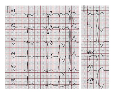
Figure 4 Electrocardiographic findings.
A 12-lead surface electrocardiogram (25 mm/s, 10 mm/mV) showing typical depolarization abnormalities (prolonged terminal activation duration in V1-V2, a minor criterion according to 2010 task force criteria, long arrows) and repolarization abnormalities (T-wave inversions V1-V4 in the absence of complete right bundle branch block, a major criterion according to 2010 task force criteria, arrowheads), and premature ventricular contractions with two different morphologies (short arrows).
- Citation: Saguner AM, Brunckhorst C, Duru F. Arrhythmogenic ventricular cardiomyopathy: A paradigm shift from right to biventricular disease. World J Cardiol 2014; 6(4): 154-174
- URL: https://www.wjgnet.com/1949-8462/full/v6/i4/154.htm
- DOI: https://dx.doi.org/10.4330/wjc.v6.i4.154


