Copyright
©2014 Baishideng Publishing Group Inc.
World J Cardiol. Nov 26, 2014; 6(11): 1166-1174
Published online Nov 26, 2014. doi: 10.4330/wjc.v6.i11.1166
Published online Nov 26, 2014. doi: 10.4330/wjc.v6.i11.1166
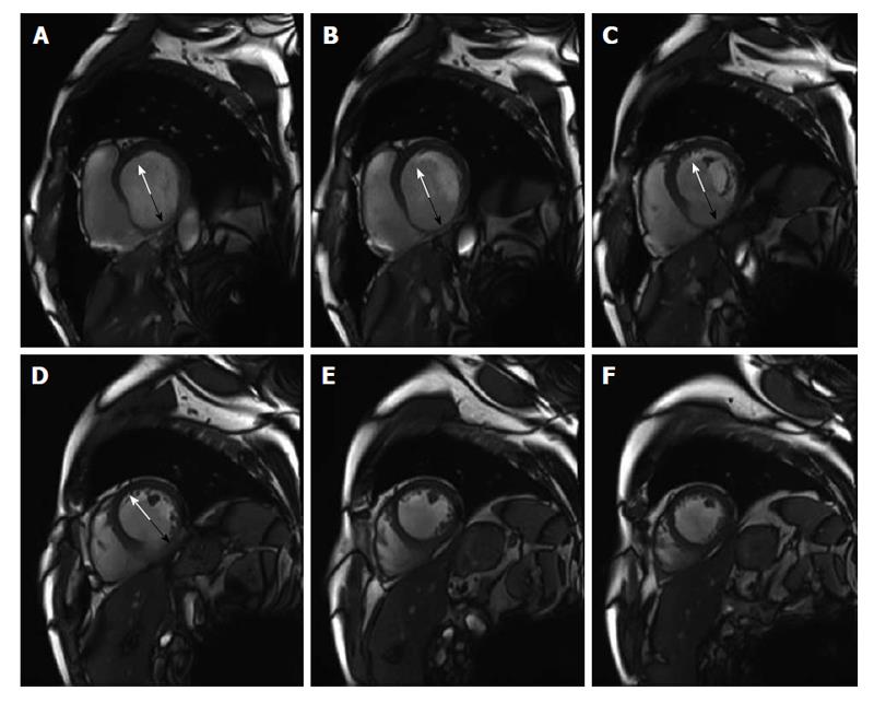
Figure 5 End diastolic wall thickness.
Representative end diastolic short axis images from basal (A) to apical (F) of a patient with previous inferior myocardial infarction. The anterior, septal and lateral region (white arrow, A-D) show a preserved end diastolic wall thickness (EDWT) > 6 mm suggesting viable myocardium, whereas EDWT of the inferior wall (black arrow, A-D) is ≤ 6 mm indicating myocardial scarring.
- Citation: Doesch C, Papavassiliu T. Diagnosis and management of ischemic cardiomyopathy: Role of cardiovascular magnetic resonance imaging. World J Cardiol 2014; 6(11): 1166-1174
- URL: https://www.wjgnet.com/1949-8462/full/v6/i11/1166.htm
- DOI: https://dx.doi.org/10.4330/wjc.v6.i11.1166


