Copyright
©2014 Baishideng Publishing Group Inc.
World J Cardiol. Nov 26, 2014; 6(11): 1166-1174
Published online Nov 26, 2014. doi: 10.4330/wjc.v6.i11.1166
Published online Nov 26, 2014. doi: 10.4330/wjc.v6.i11.1166
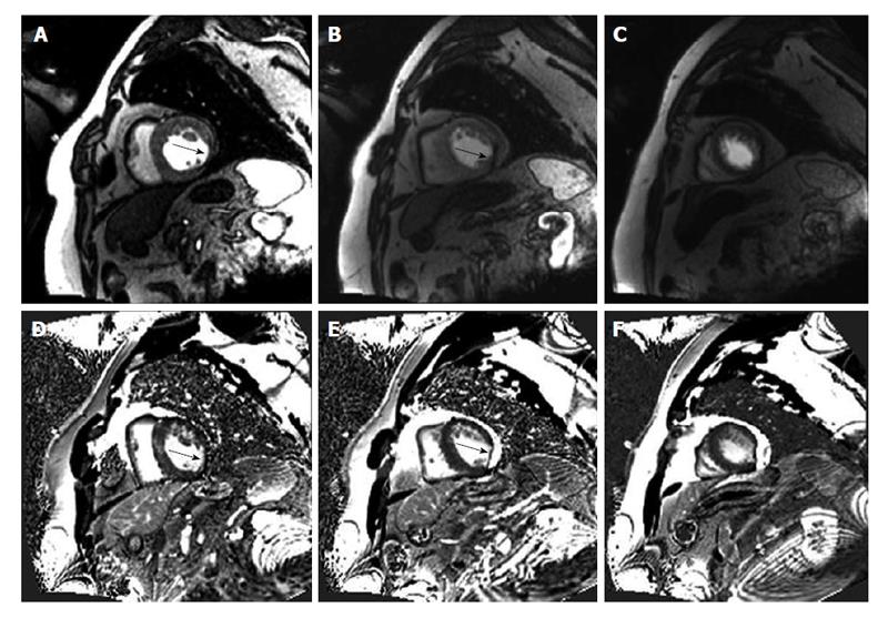
Figure 1 Patient presenting with an subacute non-ST-segment elevation infarction.
Cardiovascular magnetic resonance (CMR) images of a 54-year-old man who presented with typical chest pain. Troponin was elevated to 1.9 μg/L. CMR rest perfusion (A-C) shows a subendocardial perfusion deficit inferolateral and lateral on the basal (A) and midventricular (B) short axis slice. The black arrow highlights the subendocardial perfusion deficit. Late gadolinium enhancement (D-F) of the representative short axis also revealed a hyperenhancement inferolateral and lateral (black arrow) indicative of a subacute myocardial infarction.
- Citation: Doesch C, Papavassiliu T. Diagnosis and management of ischemic cardiomyopathy: Role of cardiovascular magnetic resonance imaging. World J Cardiol 2014; 6(11): 1166-1174
- URL: https://www.wjgnet.com/1949-8462/full/v6/i11/1166.htm
- DOI: https://dx.doi.org/10.4330/wjc.v6.i11.1166


