Copyright
©2013 Baishideng Publishing Group Co.
World J Cardiol. Sep 26, 2013; 5(9): 329-336
Published online Sep 26, 2013. doi: 10.4330/wjc.v5.i9.329
Published online Sep 26, 2013. doi: 10.4330/wjc.v5.i9.329
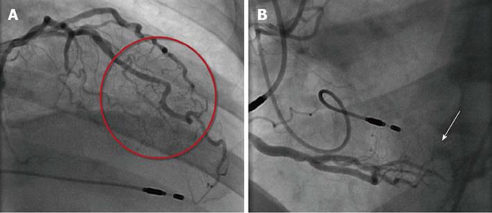
Figure 1 From the distal segment.
A: The left anterior descending coronary artery/diagonal branch multiple micro-fistulas (red circle) to the left ventricle (LV) lumen are visible; B: The right coronary artery multiple fistulas (arrow) to the LV cavity. Dual endocardial pacing leads are appreciated.
- Citation: Said SA, Schiphorst RH, Derksen R, Wagenaar L. Coronary-cameral fistulas in adults (first of two parts). World J Cardiol 2013; 5(9): 329-336
- URL: https://www.wjgnet.com/1949-8462/full/v5/i9/329.htm
- DOI: https://dx.doi.org/10.4330/wjc.v5.i9.329


