Copyright
©2013 Baishideng Publishing Group Co.
World J Cardiol. Jul 26, 2013; 5(7): 258-260
Published online Jul 26, 2013. doi: 10.4330/wjc.v5.i7.258
Published online Jul 26, 2013. doi: 10.4330/wjc.v5.i7.258
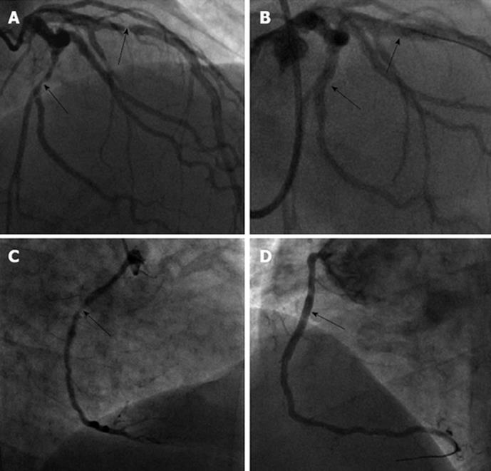
Figure 2 Coronary angiogram image.
A: Lesions in left anterior descending (LAD), left circumflex (LCx). Coronary angiogram image showing severe lesions in LAD and LCx arteries (arrows); B: Percutaneous coronary intervention to LAD and LCx. Post-percutaneous coronary intervention (PCI) images showing successful PCI to lesions in LAD and LCx (arrows); C: Lesion in right coronary artery (RCA). Coronary angiogram image showing tight lesion in RCA (arrow); D: PCI to RCA. Successful PCI to RCA with a DES (arrow).
- Citation: Hamid T, Choudhury TR, Fraser D. Multi-vessel percutaneous coronary intervention in a patient with a type B aortic dissection-transradial or transfemoral? World J Cardiol 2013; 5(7): 258-260
- URL: https://www.wjgnet.com/1949-8462/full/v5/i7/258.htm
- DOI: https://dx.doi.org/10.4330/wjc.v5.i7.258


