Copyright
©2013 Baishideng Publishing Group Co.
World J Cardiol. Jul 26, 2013; 5(7): 228-241
Published online Jul 26, 2013. doi: 10.4330/wjc.v5.i7.228
Published online Jul 26, 2013. doi: 10.4330/wjc.v5.i7.228
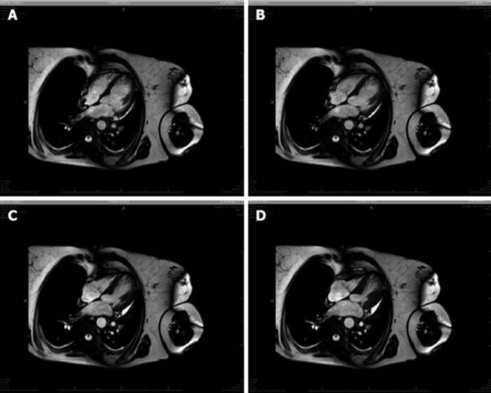
Figure 3 Cardiac magnetic resonance imaging for Takotsubo cardiomyopathy.
A: Diastole: both ventricles are distended and full of blood; B and C: Systole: both ventricles contracting; D: End of systole: the right ventricle shows a normal pattern, while the left ventricle has a ballooning shape.
- Citation: Sanchez-Jimenez EF. Initial clinical presentation of Takotsubo cardiomyopathy with-a focus on electrocardiographic changes: A literature review of cases. World J Cardiol 2013; 5(7): 228-241
- URL: https://www.wjgnet.com/1949-8462/full/v5/i7/228.htm
- DOI: https://dx.doi.org/10.4330/wjc.v5.i7.228


