Copyright
©2013 Baishideng Publishing Group Co.
World J Cardiol. May 26, 2013; 5(5): 154-156
Published online May 26, 2013. doi: 10.4330/wjc.v5.i5.154
Published online May 26, 2013. doi: 10.4330/wjc.v5.i5.154
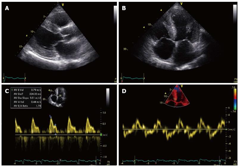
Figure 1 Transthoracic echocardiography images.
A: Parasternal long axis view diastolic still frame demonstrating thickened myocardium with sparkling of the septum. IVSd 19 mm, LPWd 19 mm; B: Apical four chamber view end diastolic still frame demonstrating thickened myocardium and normal appearance of heart valves; C: PW Doppler measurement of MV inflow. MV E/A ratio 1.8; E-vel 0.80; A-vel 0.57; IVRT 77 ms; dt 224 ms; D: Tissue Doppler Imaging with PW Doppler measurement on medial annulus of MV with E’ 3 cm/s E/E’ ratio 26.2 confirming the diastolic dysfunction. S’ 3.5 cm/s associated with impaired left ventricular function.
- Citation: Brugts JJ, Houtgraaf J, Hazenberg BP, Kofflard MJ. Echocardiographic features of an atypical presentation of rapidly progressive cardiac amyloidosis. World J Cardiol 2013; 5(5): 154-156
- URL: https://www.wjgnet.com/1949-8462/full/v5/i5/154.htm
- DOI: https://dx.doi.org/10.4330/wjc.v5.i5.154


