Copyright
©2013 Baishideng Publishing Group Co.
World J Cardiol. May 26, 2013; 5(5): 151-153
Published online May 26, 2013. doi: 10.4330/wjc.v5.i5.151
Published online May 26, 2013. doi: 10.4330/wjc.v5.i5.151
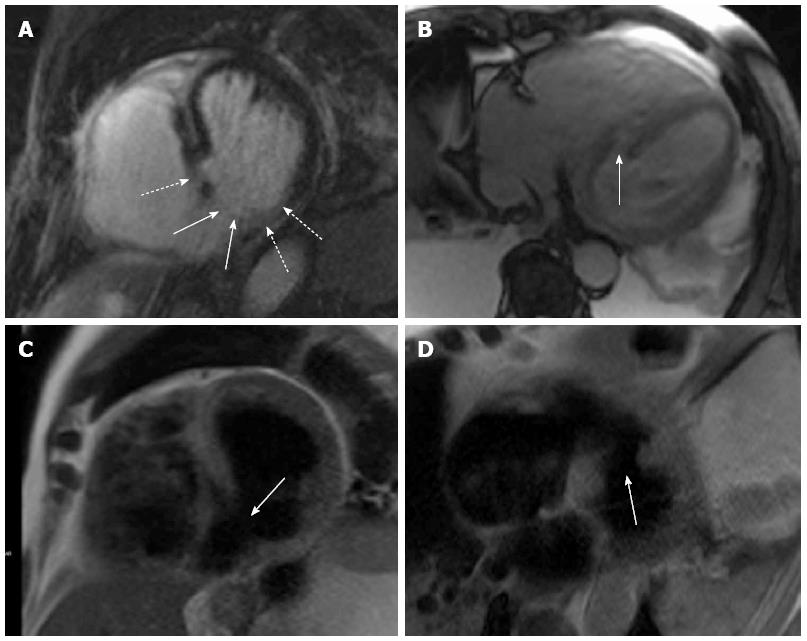
Figure 1 Cardiac magnet resonance imaging revealed size and localization of the ruptured septum with respect to the myocardial infarction zone.
Ventricular septal rupture in the infarction zone. A: Late Gadolinium enhancement in short axis view; B: On TRUFI localizer; C: On HASTE in short axis view; D: Long axis view. Solid arrows indicate ventricular septal rupture, dashed arrows indicate myocardial infarction zone.
- Citation: Gassenmaier T, Gorski A, Aleksic I, Deubner N, Weidemann F, Beer M. Impact of cardiac magnet resonance imaging on management of ventricular septal rupture after acute myocardial infarction. World J Cardiol 2013; 5(5): 151-153
- URL: https://www.wjgnet.com/1949-8462/full/v5/i5/151.htm
- DOI: https://dx.doi.org/10.4330/wjc.v5.i5.151


