Copyright
©2013 Baishideng Publishing Group Co.
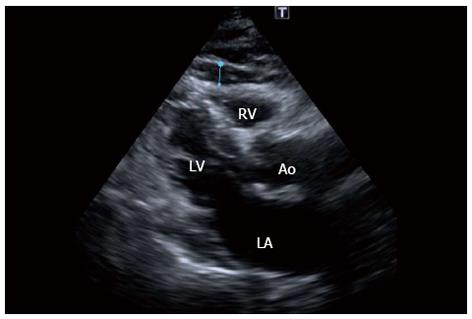
Figure 5 Echocardiogram in the parasternal long-axis view revealing a large amount of epicardial fat (thickness of 6 mm) over the free wall of the right ventricle (blue line) of the patient in figure 1.
RV: Right ventricle, LV: Left ventricle, LA: Left atrium; Ao: Aortic root.
- Citation: Echavarría-Pinto M, Hernando L, Alfonso F. From the epicardial adipose tissue to vulnerable coronary plaques. World J Cardiol 2013; 5(4): 68-74
- URL: https://www.wjgnet.com/1949-8462/full/v5/i4/68.htm
- DOI: https://dx.doi.org/10.4330/wjc.v5.i4.68


