Copyright
©2013 Baishideng Publishing Group Co.
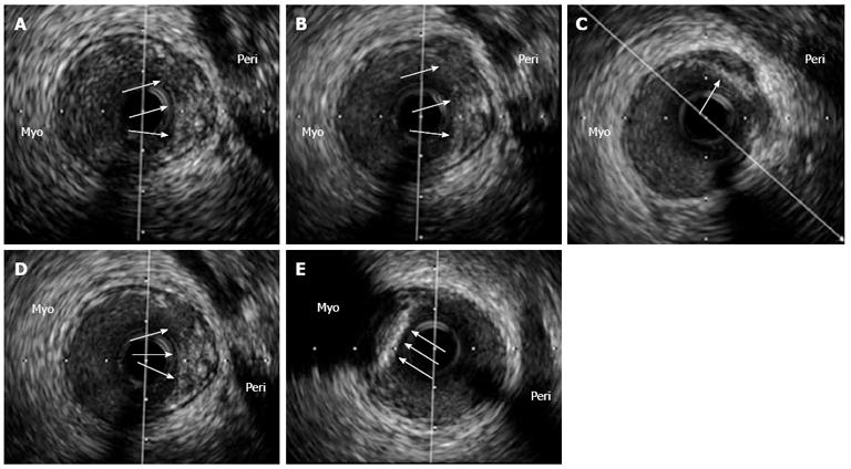
Figure 4 An intravascular ultrasound analysis of the left anterior descending coronary artery in an asymptomatic patient with a previously deployed stent (not shown) that underwent this study as part of an institutional protocol.
A-D: In the pericardial side (Peri) of the vessel, an eccentric and positively remodeled plaque with heterogenic echo-reflection that included a hypoechogenic area, suggestive of a potentially “high risk” plaque, was found (arrows); E: Interestingly, just adjacently and in the opposite vessel side [myocardial side (Myo)], a calcified plaque with intense posterior shadowing, highly suggestive of a stable fibro-calcific plaque (arrows), was also observed. The patient has been asymptomatic during a 3-year clinical follow-up.
- Citation: Echavarría-Pinto M, Hernando L, Alfonso F. From the epicardial adipose tissue to vulnerable coronary plaques. World J Cardiol 2013; 5(4): 68-74
- URL: https://www.wjgnet.com/1949-8462/full/v5/i4/68.htm
- DOI: https://dx.doi.org/10.4330/wjc.v5.i4.68


