Copyright
©2013 Baishideng Publishing Group Co.
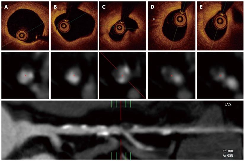
Figure 3 Optical coherence tomography and multislice computed tomography findings of the same patient in figure 2.
A and B: In the upper panel, optical coherence tomography revealed a complex plaque affecting a long portion of the vessel with calcified and lipidic regions; C: Also, a complex fibroatheroma that included a lipidic core was observed at the site of the most severe stenosis; D: A high light attenuation band with distal shadowing can be attributed to macrophage infiltration (*). The middle panel represents cross-sectional views of the vessel at the same stenosis as visualized by multislice computed tomography (MSCT). Calcified and non-calcified regions including eccentric plaques were found within the diseased segment. The inferior panel shows a MSCT multiplanar reconstruction of the vessel and its anatomical bookmarks.
- Citation: Echavarría-Pinto M, Hernando L, Alfonso F. From the epicardial adipose tissue to vulnerable coronary plaques. World J Cardiol 2013; 5(4): 68-74
- URL: https://www.wjgnet.com/1949-8462/full/v5/i4/68.htm
- DOI: https://dx.doi.org/10.4330/wjc.v5.i4.68


