Copyright
©2013 Baishideng Publishing Group Co.
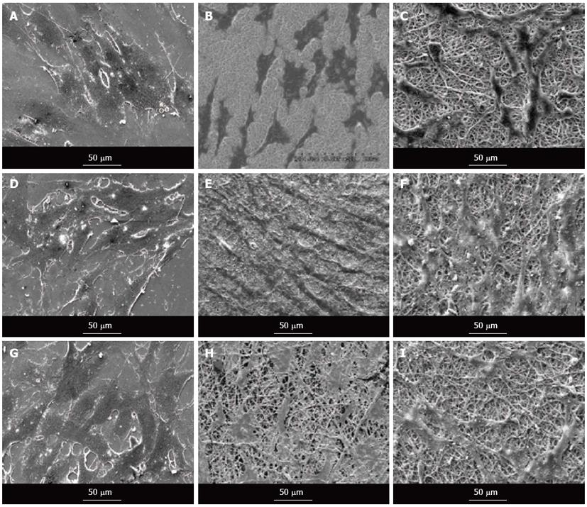
Figure 9 Scanning electron microscope images showing the cell morphology of cardiomyocytes (A-C), cardiomyocytes-mesenchymal stem cells co-culture cells (D-F) and mesenchymal stem cells (G-I) grown on tissue culture plate (A, D, G), collagen nanofibers (B, E, H) and poly (glycerol sebacate)/collagen core/shell fibers (C, F, I) on day 15 at 500 × magnification.
- Citation: Ravichandran R, Venugopal JR, Sundarrajan S, Mukherjee S, Ramakrishna S. Cardiogenic differentiation of mesenchymal stem cells on elastomeric poly (glycerol sebacate)/collagen core/shell fibers. World J Cardiol 2013; 5(3): 28-41
- URL: https://www.wjgnet.com/1949-8462/full/v5/i3/28.htm
- DOI: https://dx.doi.org/10.4330/wjc.v5.i3.28


