Copyright
©2013 Baishideng.
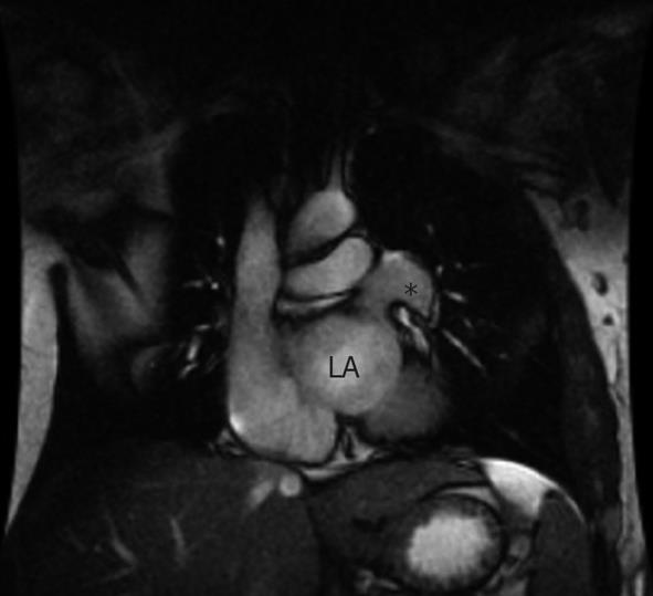
Figure 2 Cardiac magnetic resonance imaging.
Cardiac magnetic resonance imaging (coronal view) displaying the partial pericardial defect (20 mm × 30 mm) localized to the left atrial (LA) wall. Herniation of the left atrial appendage can been seen (asterisk).
- Citation: Juárez AL, Akerström F, Alguacil AM, González BS. Congenital partial absence of the pericardium in a young man with atypical chest pain. World J Cardiol 2013; 5(2): 12-14
- URL: https://www.wjgnet.com/1949-8462/full/v5/i2/12.htm
- DOI: https://dx.doi.org/10.4330/wjc.v5.i2.12


