Copyright
©2013 Baishideng Publishing Group Co.
World J Cardiol. Nov 26, 2013; 5(11): 397-403
Published online Nov 26, 2013. doi: 10.4330/wjc.v5.i11.397
Published online Nov 26, 2013. doi: 10.4330/wjc.v5.i11.397
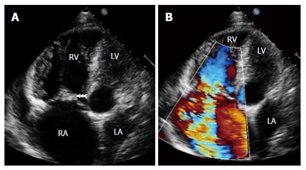
Figure 2 Transthoracic echocardiography showing (A) large vegetation attached to tricuspid valve leaflets (arrowheads) in a patient with intravenous drug abuse and septic pulmonary emboli.
Note hugely dilated right atrium and right ventricle and (B) severe tricuspid regurgitation. RA: Right atrium; RV: Right ventricle; LA: Left atrium; LV: Left ventricle.
- Citation: Panduranga P, Al-Abri S, Al-Lawati J. Intravenous drug abuse and tricuspid valve endocarditis: Growing trends in the Middle East Gulf region. World J Cardiol 2013; 5(11): 397-403
- URL: https://www.wjgnet.com/1949-8462/full/v5/i11/397.htm
- DOI: https://dx.doi.org/10.4330/wjc.v5.i11.397


