Copyright
©2012 Baishideng Publishing Group Co.
World J Cardiol. May 26, 2012; 4(5): 188-194
Published online May 26, 2012. doi: 10.4330/wjc.v4.i5.188
Published online May 26, 2012. doi: 10.4330/wjc.v4.i5.188
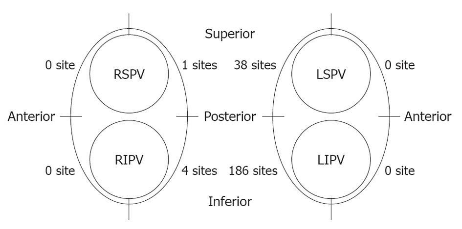
Figure 3 Position of sites where luminal esophageal temperature reached the cut-off temperature.
Most of the sites were located along the posterior side of the LPV, especially around the left inferior pulmonary vein (LIPV), while only five sites were observed near the RPVs. LSPV: Left superior pulmonary vein; RSPV: Right superior pulmonary vein; RIPV: Right inferior pulmonary vein.
- Citation: Sato D, Teramoto K, Kitajima H, Nishina N, Kida Y, Mani H, Esato M, Chun YH, Iwasaka T. Measuring luminal esophageal temperature during pulmonary vein isolation of atrial fibrillation. World J Cardiol 2012; 4(5): 188-194
- URL: https://www.wjgnet.com/1949-8462/full/v4/i5/188.htm
- DOI: https://dx.doi.org/10.4330/wjc.v4.i5.188


