Copyright
©2012 Baishideng Publishing Group Co.
World J Cardiol. May 26, 2012; 4(5): 173-182
Published online May 26, 2012. doi: 10.4330/wjc.v4.i5.173
Published online May 26, 2012. doi: 10.4330/wjc.v4.i5.173
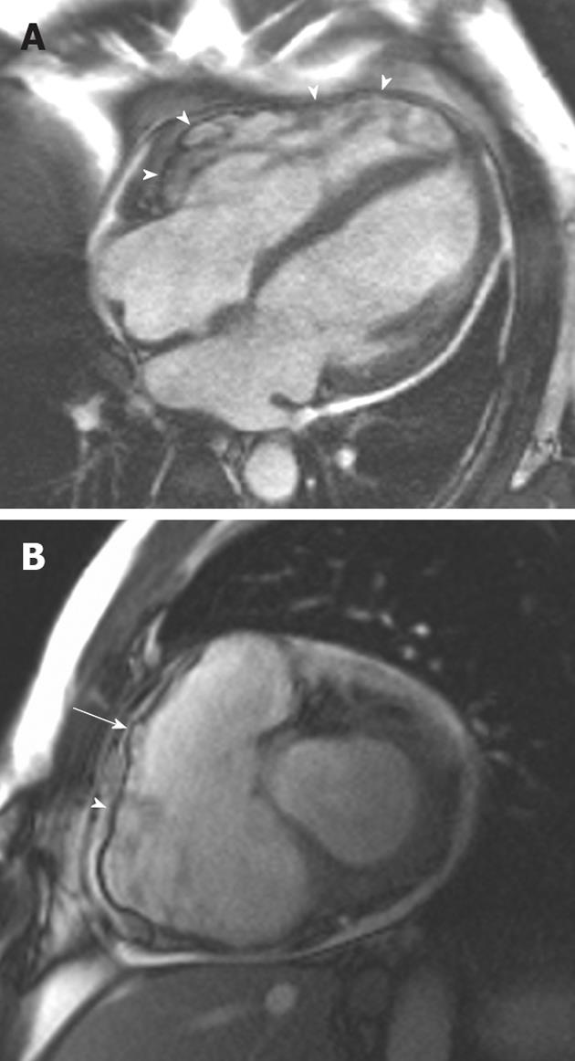
Figure 3 A 38-year-old man of Italian origin presented with palpitations and progressive shortness of breath during exercise.
A: Horizontal long-axis SSFP shows a heavily trabeculated right ventricle with thickened trabeculae and increased right ventricular (RV) volume. The RV free wall is very thin, with multiple small aneurysms (arrowheads); B: Short-axis SSFP sequence showing further small aneurysms of the RV free wall. Note again the increased RV volume.
- Citation: O’Neill AC, McDermott S, Ridge CA, Keane D, Dodd JD. Investigation of cardiomyopathy using cardiac magnetic resonance imaging part 2: Rare phenotypes. World J Cardiol 2012; 4(5): 173-182
- URL: https://www.wjgnet.com/1949-8462/full/v4/i5/173.htm
- DOI: https://dx.doi.org/10.4330/wjc.v4.i5.173


