Copyright
©2012 Baishideng Publishing Group Co.
World J Cardiol. Apr 26, 2012; 4(4): 103-111
Published online Apr 26, 2012. doi: 10.4330/wjc.v4.i4.103
Published online Apr 26, 2012. doi: 10.4330/wjc.v4.i4.103
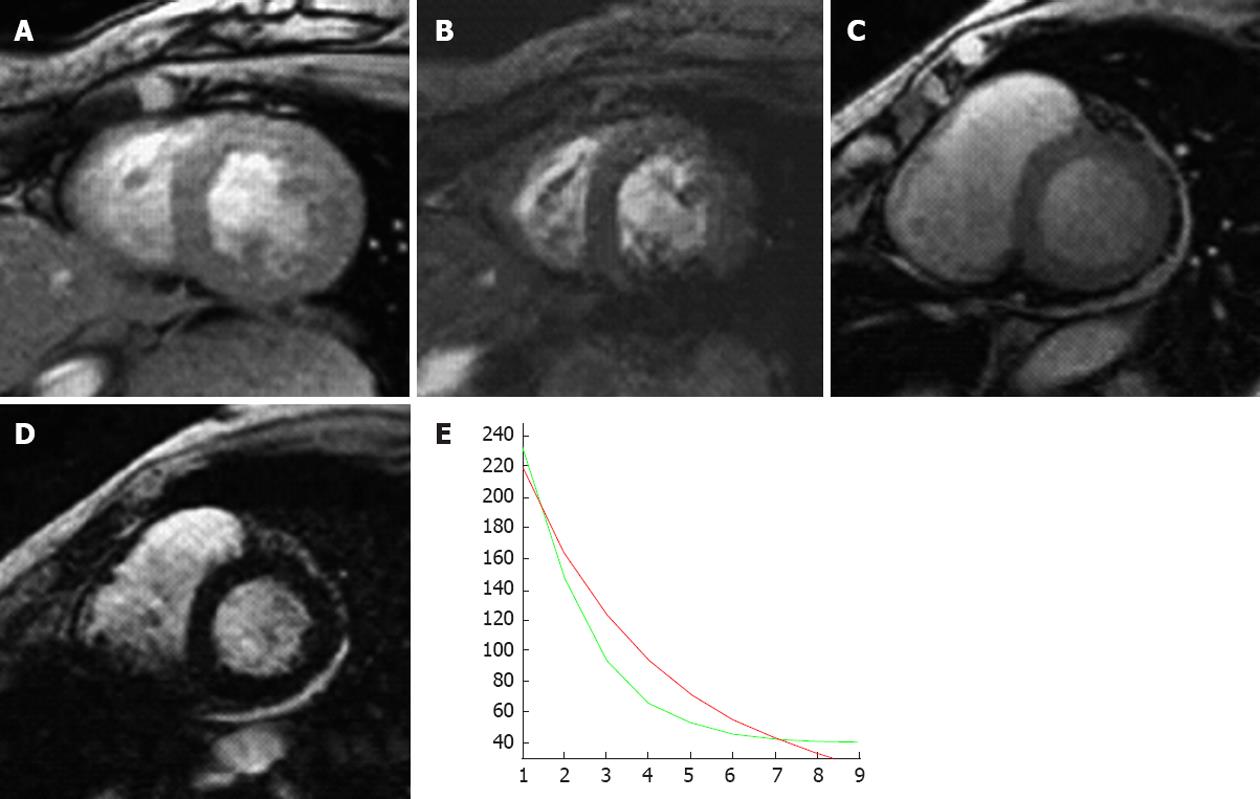
Figure 6 A 79-year-old woman with myelodysplasia treated with blood transfusions over many years.
A (echo time 10 ms) and B (echo time 20 ms) are two short axis T2star sequences from a normal patient with no evidence of myocardial iron overload; C (echo time 10 ms) and D (echo time 20 ms) are from the patient with myelodysplasia showing a progressive loss of signal with increasing T2 echo time indicating shortened T1 relation secondary to iron infiltration; E: Graph of decreasing T1 relaxation times (green line) compared with a normal patient (red line).
- Citation: McDermott S, O’Neill AC, Ridge CA, Dodd JD. Investigation of cardiomyopathy using cardiac magnetic resonance imaging part 1: Common phenotypes. World J Cardiol 2012; 4(4): 103-111
- URL: https://www.wjgnet.com/1949-8462/full/v4/i4/103.htm
- DOI: https://dx.doi.org/10.4330/wjc.v4.i4.103


