Copyright
©2012 Baishideng Publishing Group Co.
World J Cardiol. Apr 26, 2012; 4(4): 103-111
Published online Apr 26, 2012. doi: 10.4330/wjc.v4.i4.103
Published online Apr 26, 2012. doi: 10.4330/wjc.v4.i4.103
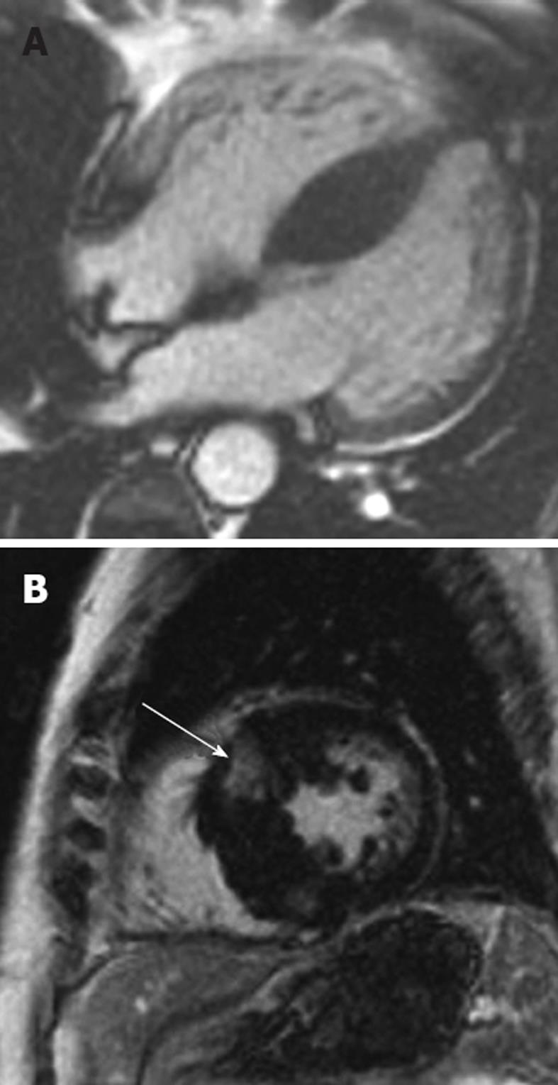
Figure 2 Hypertrophic cardiomyopathy.
A: 28-year-old man who presented with progressive heart failure and palpitations. The horizontal long-axis steady-state free precession sequence demonstrates a hypertrophic interventricular septum measuring 23 mm (normal ≤ 11 mm) consistent with hypertrophic cardiomyopathy (HCM); B: Late-enhancement short-axis image shows late-enhancement in the hypertrophied septum (arrow). Note that there are 2 abnormal areas of enhancement corresponding to the right superior and inferior ventricular insertion points. This is a characteristic pattern in HCM. Such late-enhancement has prognostic implications for patients with HCM, being associated with an increased prevalence of heart failure admissions, deterioration to New York Heart Association functional class III or IV, or heart failure-related death.
- Citation: McDermott S, O’Neill AC, Ridge CA, Dodd JD. Investigation of cardiomyopathy using cardiac magnetic resonance imaging part 1: Common phenotypes. World J Cardiol 2012; 4(4): 103-111
- URL: https://www.wjgnet.com/1949-8462/full/v4/i4/103.htm
- DOI: https://dx.doi.org/10.4330/wjc.v4.i4.103


