Copyright
©2012 Baishideng Publishing Group Co.
World J Cardiol. Nov 26, 2012; 4(11): 309-311
Published online Nov 26, 2012. doi: 10.4330/wjc.v4.i11.309
Published online Nov 26, 2012. doi: 10.4330/wjc.v4.i11.309
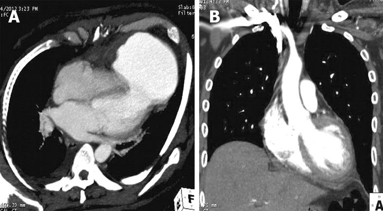
Figure 2 Computed tomography image of the heart.
A: Computed tomography (CT) angiographic oblique axial image shows a rent of 28 mm at the left ventricle (LV) apex, communicating with a large 76 mm × 98 mm pseudoaneurysm; B: Post surgery, CT angiographic oblique coronal image in the plane of LV outflow tract shows well-delineated LV cavity without any pseudoaneurysm.
- Citation: Vijayvergiya R, Kumar A, Rana SS, Singh H, Puri GD, Singhal M. Post-myocardial infarction giant left ventricular pseudoaneurysm presenting with severe heart failure. World J Cardiol 2012; 4(11): 309-311
- URL: https://www.wjgnet.com/1949-8462/full/v4/i11/309.htm
- DOI: https://dx.doi.org/10.4330/wjc.v4.i11.309


