Copyright
©2012 Baishideng Publishing Group Co.
World J Cardiol. Oct 26, 2012; 4(10): 296-301
Published online Oct 26, 2012. doi: 10.4330/wjc.v4.i10.296
Published online Oct 26, 2012. doi: 10.4330/wjc.v4.i10.296
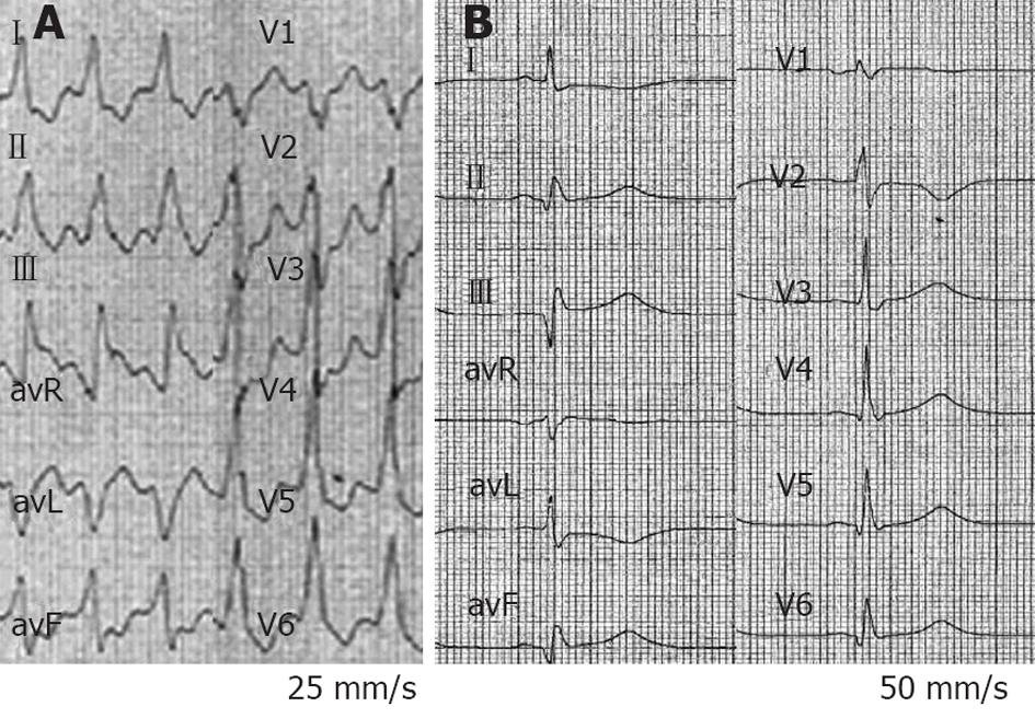
Figure 4 Electrocardiogram tracings from a 64-year-old man with ischemic dilated cardiomyopathy with an implanted cardioverter-defibrillator, on chronic treatment with bisoprolol and amiodarone (patient 2).
A: Ten-lead electrocardiogram (ECG) at admission with sustained ventricular tachycardia (VT) at 140 beats/min with a left bundle branch block morphology and right axis deviation, blood pressure was 85/50 mmHg; B: Twelve-lead ECG 30 s after intravenous epinephrine bolus (0.5 mg over 30 s) shows sinus rhythm with pathologic Q waves in inferior leads. VT termination was preceded by a slight increase in VT rate from 140 to 148 beats/min (not shown).
- Citation: Bonny A, De Sisti A, Márquez MF, Megbemado R, Hidden-Lucet F, Fontaine G. Low doses of intravenous epinephrine for refractory sustained monomorphic ventricular tachycardia. World J Cardiol 2012; 4(10): 296-301
- URL: https://www.wjgnet.com/1949-8462/full/v4/i10/296.htm
- DOI: https://dx.doi.org/10.4330/wjc.v4.i10.296


