Copyright
©2012 Baishideng Publishing Group Co.
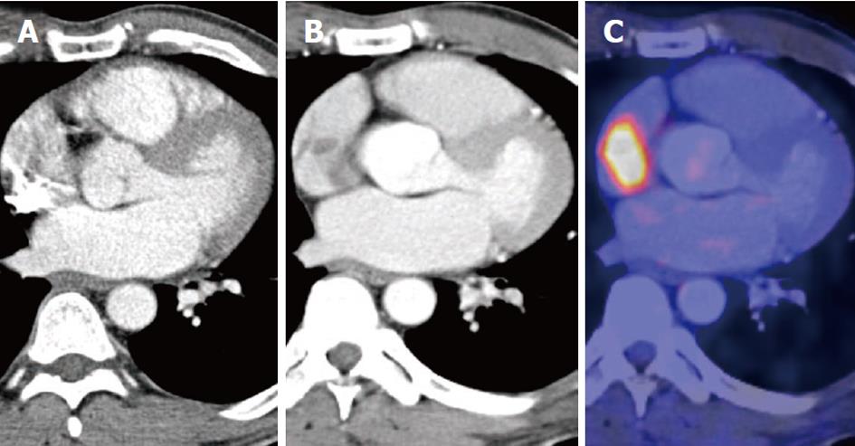
Figure 1 Computed tomography scan.
A: Initial computed tomography (CT) scan, with artifacts in the right atrium; B: CT of better quality; C: 18F-fluorodeoxyglucose positron emission tomography-CT with specific tracer accumulation.
- Citation: Krüger T, Heuschmid M, Kurth R, Stock UA, Wildhirt SM. Asymptomatic melanoma of the superior cavo-atrial junction: The challenge of imaging. World J Cardiol 2012; 4(1): 20-22
- URL: https://www.wjgnet.com/1949-8462/full/v4/i1/20.htm
- DOI: https://dx.doi.org/10.4330/wjc.v4.i1.20


