Copyright
©2011 Baishideng Publishing Group Co.
World J Cardiol. Nov 26, 2011; 3(11): 339-350
Published online Nov 26, 2011. doi: 10.4330/wjc.v3.i11.339
Published online Nov 26, 2011. doi: 10.4330/wjc.v3.i11.339
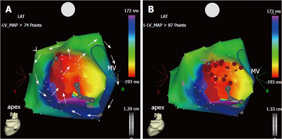
Figure 2 Three-dimensional electroanatomic map during macroreentrant ventricular tachycardia (A, B).
Postero-anterior view of the electroanatomic activation map of the left ventricular endocardium, reconstructed during ventricular tachycardia with a cycle length of 380 ms in a patient with postinfarction ischemic cardiomyopathy. A: The ventricular tachycardia is sustained by a reentry circuit, which has been fully reconstructed during point-by-point mapping. The reentry course is shown by the sequence of colors from red to purple (arrows). There are two reentrant loops: one rotates clockwise around the mitral annulus, whereas the other has a counter-clockwise course in the lateral wall of the left ventricle. Both loops share the mid-diastolic isthmus, the dark red area between two electrically silent areas (gray dots) where the two arrowed circles meet each other; B: Sequential radiofrequency energy applications (red dots) were delivered linearly to transect the critical isthmus and produce a line of block between the two electrically silent areas and the mitral annulus. No arrhythmia was inducible at the end of the procedure and the patient had no recurrence at the mid-term follow-up. MV: Mitral valve.
- Citation: Ponti RD. Role of catheter ablation of ventricular tachycardia associated with structural heart disease. World J Cardiol 2011; 3(11): 339-350
- URL: https://www.wjgnet.com/1949-8462/full/v3/i11/339.htm
- DOI: https://dx.doi.org/10.4330/wjc.v3.i11.339


