Copyright
©2011 Baishideng Publishing Group Co.
World J Cardiol. Oct 26, 2011; 3(10): 337-338
Published online Oct 26, 2011. doi: 10.4330/wjc.v3.i10.337
Published online Oct 26, 2011. doi: 10.4330/wjc.v3.i10.337
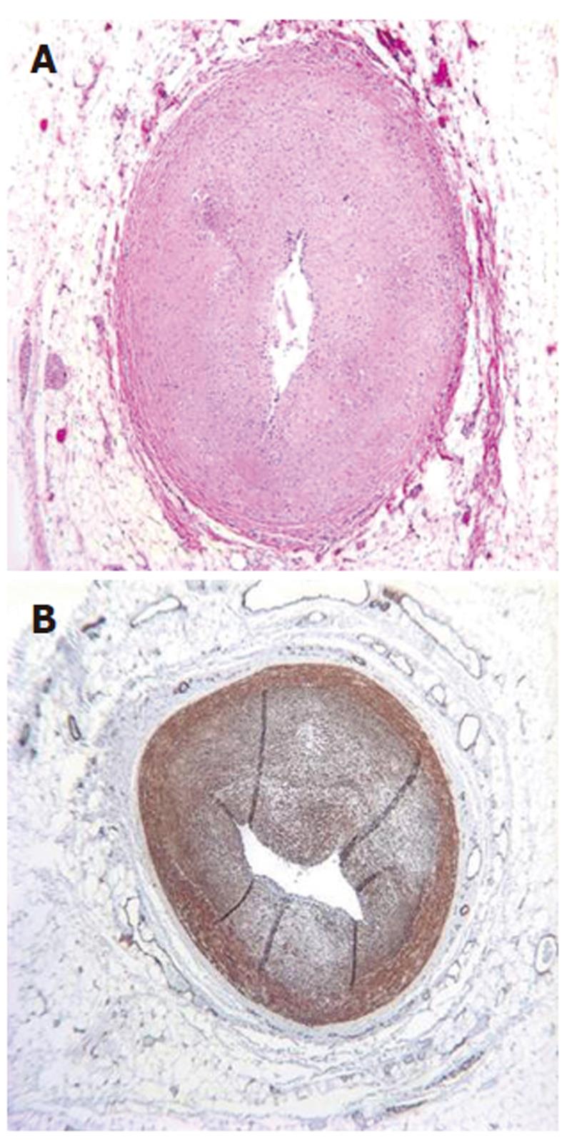
Figure 1 Sections at different segments of the proximal left anterior descending coronary artery.
A: Hematoxylin and eosin stain; B: Smooth muscle actin immunohistochemistry (Original magnification, 40 ×). Note intimal expansion and critical luminal compromise in both sections; B shows that most of the intimal expansion is due to smooth muscle cells.
- Citation: Bhavsar T, Hayes T, Wurzel J. Epicardial coronary artery intimal smooth muscle hyperplasia in a cocaine user. World J Cardiol 2011; 3(10): 337-338
- URL: https://www.wjgnet.com/1949-8462/full/v3/i10/337.htm
- DOI: https://dx.doi.org/10.4330/wjc.v3.i10.337


