Copyright
©2010 Baishideng Publishing Group Co.
World J Cardiol. Aug 26, 2010; 2(8): 223-232
Published online Aug 26, 2010. doi: 10.4330/wjc.v2.i8.223
Published online Aug 26, 2010. doi: 10.4330/wjc.v2.i8.223
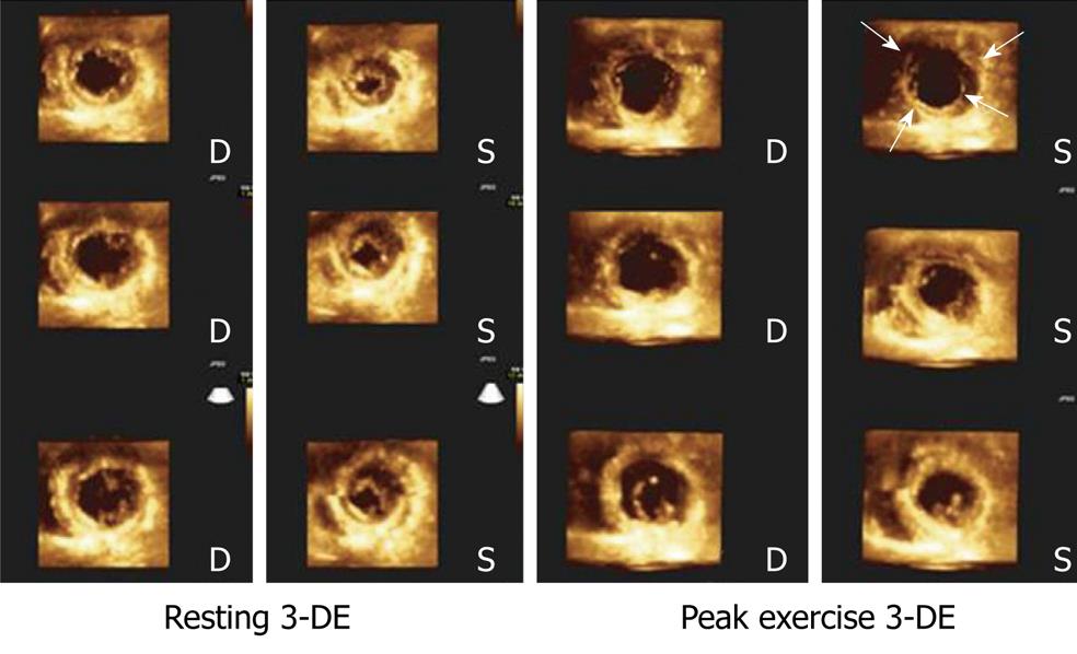
Figure 6 Cropped views obtained from a left ventricle full-volume during 3-dimensional exercise echocardiography in resting conditions (left panel) and during peak exercise (right panel) in a patient with severe 3-vessel disease.
Note exercise-induced akinesia and dilation in the short axis apical views (arrows), as well as hypokinesia and dilation in the short-axis view at the papillary muscles level. 3-DE: Three-dimensional exercise; D: Diastolic; S: Systolic.
- Citation: Peteiro J, Bouzas-Mosquera A. Exercise echocardiography. World J Cardiol 2010; 2(8): 223-232
- URL: https://www.wjgnet.com/1949-8462/full/v2/i8/223.htm
- DOI: https://dx.doi.org/10.4330/wjc.v2.i8.223


