Copyright
©2010 Baishideng Publishing Group Co.
World J Cardiol. Aug 26, 2010; 2(8): 223-232
Published online Aug 26, 2010. doi: 10.4330/wjc.v2.i8.223
Published online Aug 26, 2010. doi: 10.4330/wjc.v2.i8.223
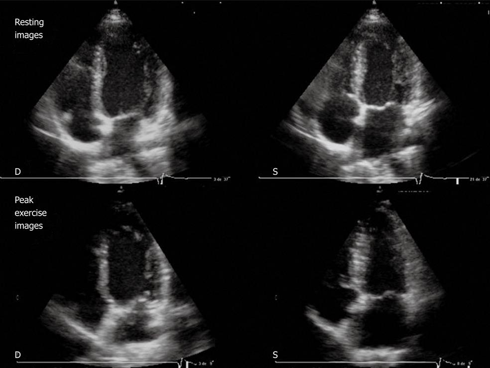
Figure 2 Resting echocardiography (top) and peak exercise echocardiography (bottom).
Four-chamber apical view (diastolic frames on the left, systolic frames on the right) in a patient with normal results. Note the left ventricular (LV) cavity dimensions decrease with exercise and an increase in LV ejection fraction. D: Diastolic; S: Systolic.
- Citation: Peteiro J, Bouzas-Mosquera A. Exercise echocardiography. World J Cardiol 2010; 2(8): 223-232
- URL: https://www.wjgnet.com/1949-8462/full/v2/i8/223.htm
- DOI: https://dx.doi.org/10.4330/wjc.v2.i8.223


