Copyright
©2010 Baishideng Publishing Group Co.
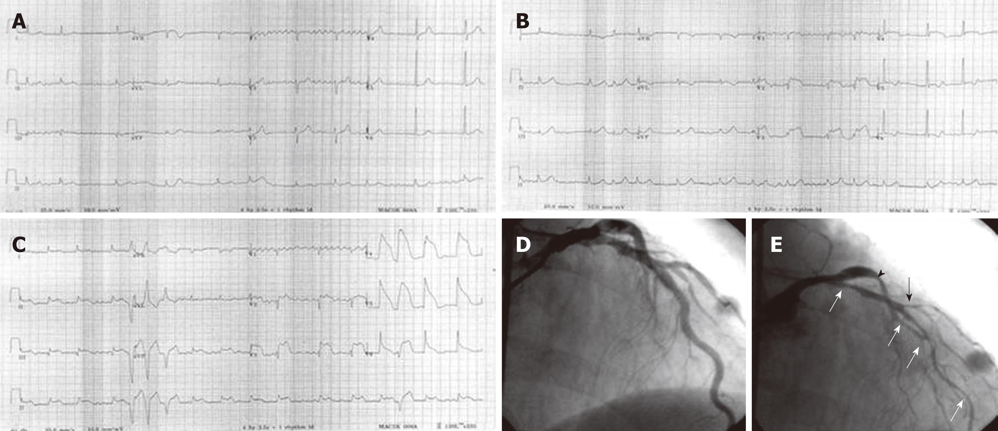
Figure 1 Baseline electrocardiography (ECG) of a patient with variant angina.
A: The ECG showed no evidence of myocardial ischemia on admission to the emergency department; B: A few hours later, the follow-up ECG, due to chest pain, showed ST-segment elevation in the V2-4 leads. C: During the following days, serial ECGs due to chest pain showed dynamic ST-segment elevation in the anterior and inferior leads. D: The patient’s ECG was normal when he was not having chest pain. Baseline coronary angiography showed no evidence of significant fixed coronary artery stenosis; E: Diffuse spasm in the proximal to distal portion (white arrows) and diagonal branch (black arrow) of the left anterior descending artery and in the proximal portion of the left circumflex artery (arrowhead) were noted following intracoronary methylergonovine administration.
- Citation: Hung MJ. Current advances in the understanding of coronary vasospasm. World J Cardiol 2010; 2(2): 34-42
- URL: https://www.wjgnet.com/1949-8462/full/v2/i2/34.htm
- DOI: https://dx.doi.org/10.4330/wjc.v2.i2.34


