Copyright
©2010 Baishideng Publishing Group Co.
World J Cardiol. Oct 26, 2010; 2(10): 333-343
Published online Oct 26, 2010. doi: 10.4330/wjc.v2.i10.333
Published online Oct 26, 2010. doi: 10.4330/wjc.v2.i10.333
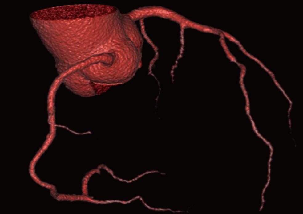
Figure 3 Three-dimensional volume rendering of the coronary arteries and side branches are clearly visualized with use of 320-slice computed tomography angiography in a 58-year-old man presenting with chest pain.
Volumetric data were acquired within a single heartbeat with excellent image quality.
- Citation: Sun Z. Multislice CT angiography in coronary artery disease: Technical developments, radiation dose and diagnostic value. World J Cardiol 2010; 2(10): 333-343
- URL: https://www.wjgnet.com/1949-8462/full/v2/i10/333.htm
- DOI: https://dx.doi.org/10.4330/wjc.v2.i10.333


