Copyright
©2010 Baishideng Publishing Group Co.
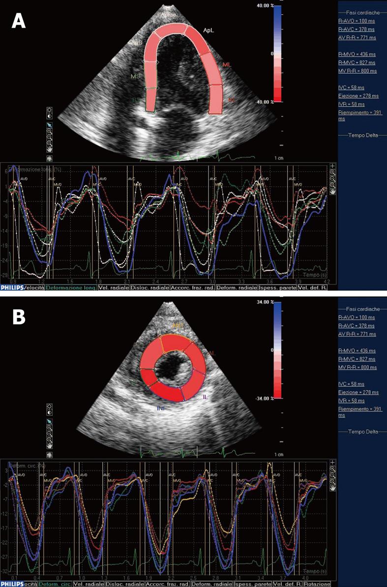
Figure 1 Systolic myocardial deformation after electro-mechanical activation.
A: LV longitudinal strain from the apical four-chamber view: time-strain curves show a negative end-systolic strain representing myocardial shortening during systole; B: LV circumferential strain from the short axis view: time-strain curves show a negative end-systolic strain representing myocardial shortening during systole. End-systole has been identified by the AVC. At this point, we could observe the negative peak of the time-strain curves corresponding to each myocardial segment.
- Citation: Sitia S, Tomasoni L, Turiel M. Speckle tracking echocardiography: A new approach to myocardial function. World J Cardiol 2010; 2(1): 1-5
- URL: https://www.wjgnet.com/1949-8462/full/v2/i1/1.htm
- DOI: https://dx.doi.org/10.4330/wjc.v2.i1.1


