Copyright
©The Author(s) 2025.
World J Cardiol. Feb 26, 2025; 17(2): 101588
Published online Feb 26, 2025. doi: 10.4330/wjc.v17.i2.101588
Published online Feb 26, 2025. doi: 10.4330/wjc.v17.i2.101588
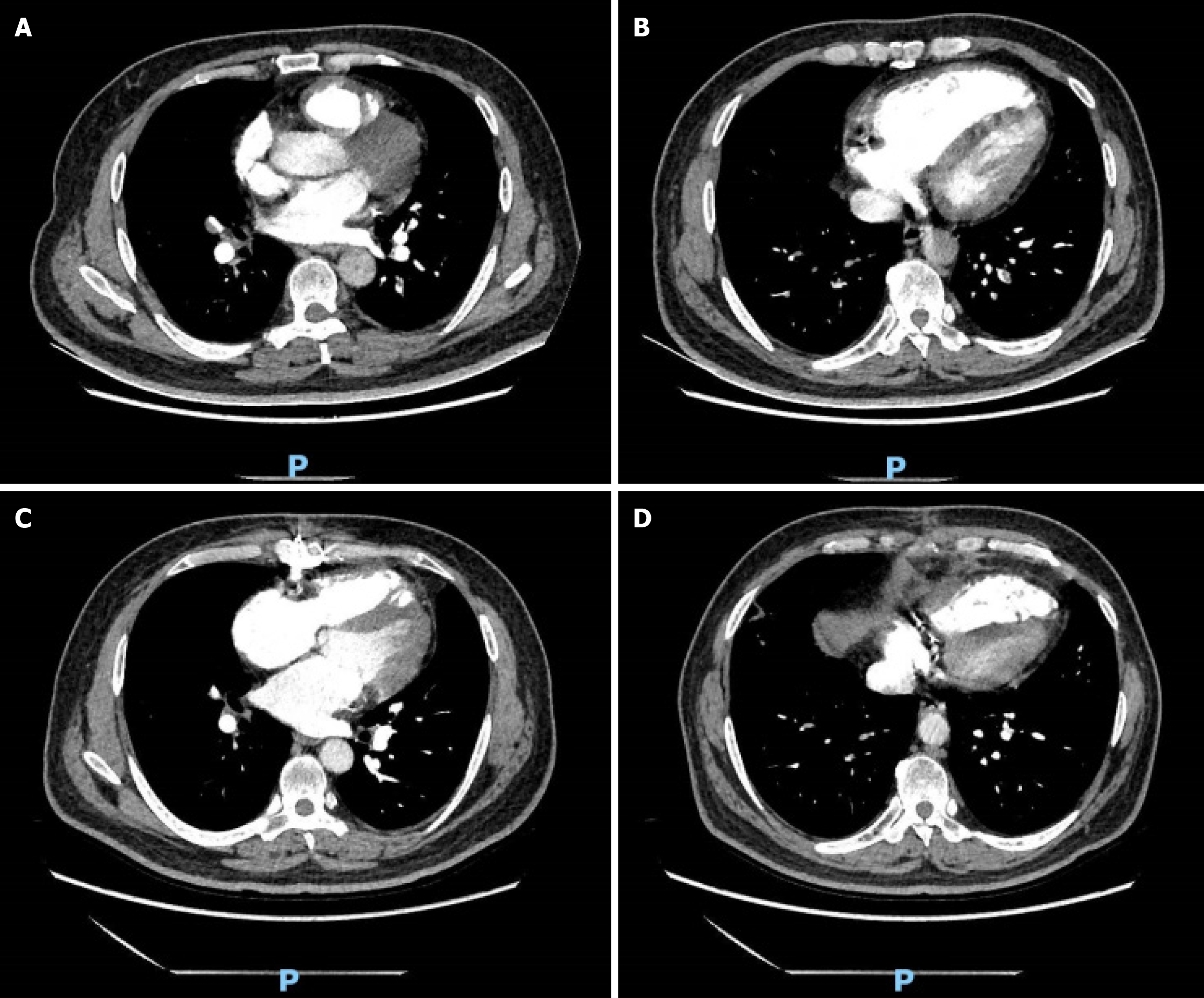
Figure 1 Changes in computed tomography pulmonary angiography after treatment.
A and B: Computed tomography pulmonary angiography (CTPA) after admission, indicating thickening of the pulmonary arterial trunk and multiple low-density filling deficiencies in the pulmonary artery in the right middle lobe and in bilateral segmental arteries; C and D: CTPA after 2 mo of follow-up, showing slight thickening of the pulmonary trunk and striated filling defects with lumen stenosis in several segmental pulmonary arteries and their branches bilaterally. Notably, a reduction in the extent of filling defects within both pulmonary arteries is observed compared to the initial CTPA.
- Citation: Xu JQ, Jiang MX, Wang F, Yang KQ, Xu YJ, Wang YJ, Dong SJ. Coronary heart disease with pulmonary embolism: A case report. World J Cardiol 2025; 17(2): 101588
- URL: https://www.wjgnet.com/1949-8462/full/v17/i2/101588.htm
- DOI: https://dx.doi.org/10.4330/wjc.v17.i2.101588


