Copyright
©The Author(s) 2025.
World J Cardiol. Feb 26, 2025; 17(2): 100952
Published online Feb 26, 2025. doi: 10.4330/wjc.v17.i2.100952
Published online Feb 26, 2025. doi: 10.4330/wjc.v17.i2.100952
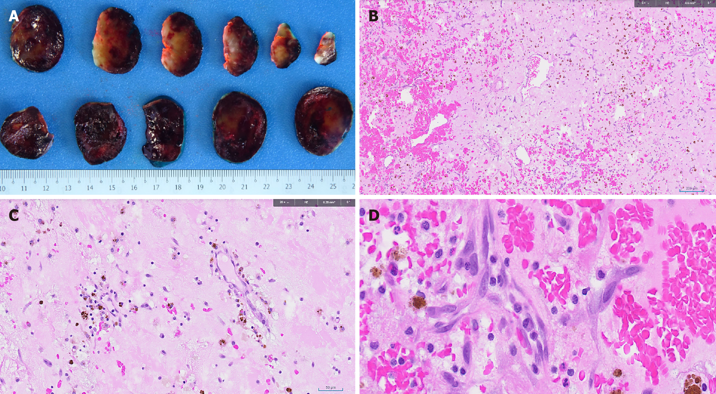
Figure 4 Histopathological findings of the left atrial tumor.
A: On macroscopic examination, cut sections show a predominantly hemorrhagic appearance with some pale yellow myxoid areas. No fleshy areas are identified (scale included at the bottom of the image); B: Low power microscopic examination shows stellate, ovoid, and plump spindle cells set in a vascularized myxoid stroma [hematoxylin and eosin-stained slide (HE) 50 × magnification]; C: The cells are singly dispersed, and arranged in perivascular rings around small blood vessels (HE-stained slide, 200 × magnification); D: There is no high-grade cytological atypia. There is prominent intratumoral hemorrhage and hemosiderin deposition. Mixed inflammatory infiltrate is noted (HE-stained slide, 400 × magnification).
- Citation: Zhu L, Neo JYZ, Punjabi LS, Lai SH, Chua YL. Large left atrial myxoma with synchronous laryngeal squamous cell carcinoma: A case report. World J Cardiol 2025; 17(2): 100952
- URL: https://www.wjgnet.com/1949-8462/full/v17/i2/100952.htm
- DOI: https://dx.doi.org/10.4330/wjc.v17.i2.100952


