Copyright
©The Author(s) 2025.
World J Cardiol. Jan 26, 2025; 17(1): 102147
Published online Jan 26, 2025. doi: 10.4330/wjc.v17.i1.102147
Published online Jan 26, 2025. doi: 10.4330/wjc.v17.i1.102147
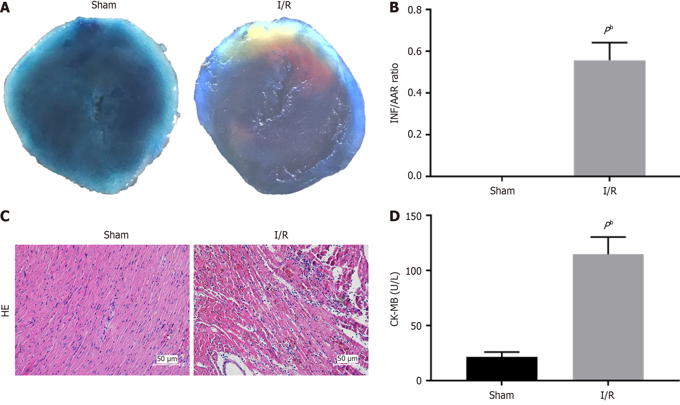
Figure 1 Ischemia/reperfusion induced myocardial injury in mice.
A: Representative images of the infract area international normalized ratio (INR) (white), area at risk (AAR) (red and white), and normal area (blue); B: Quantitative analysis of infarct size and the ratio of INF/AAR; C: Representative images of myocardial tissues from the myocardial ischemia/reperfusion group and sham group; D: Quantitative analysis of creatine kinase-myocardial bound level. All Data are shown as means ± SD. aP < 0.05, bP < 0.01 vs sham group, n = 6 per group. AAR: Area at risk; CK-MB: Creatine kinase-myocardial bound; HE: Hematoxylin-eosin; INR: International normalized ratio; I/R: Ischemia/reperfusion.
- Citation: Wang JN, Zhou YY, Yu YW, Chen J. Profiling and bioinformatics analyses of circular RNAs in myocardial ischemia/reperfusion injury model in mice. World J Cardiol 2025; 17(1): 102147
- URL: https://www.wjgnet.com/1949-8462/full/v17/i1/102147.htm
- DOI: https://dx.doi.org/10.4330/wjc.v17.i1.102147


