Copyright
©The Author(s) 2024.
World J Cardiol. Sep 26, 2024; 16(9): 531-541
Published online Sep 26, 2024. doi: 10.4330/wjc.v16.i9.531
Published online Sep 26, 2024. doi: 10.4330/wjc.v16.i9.531
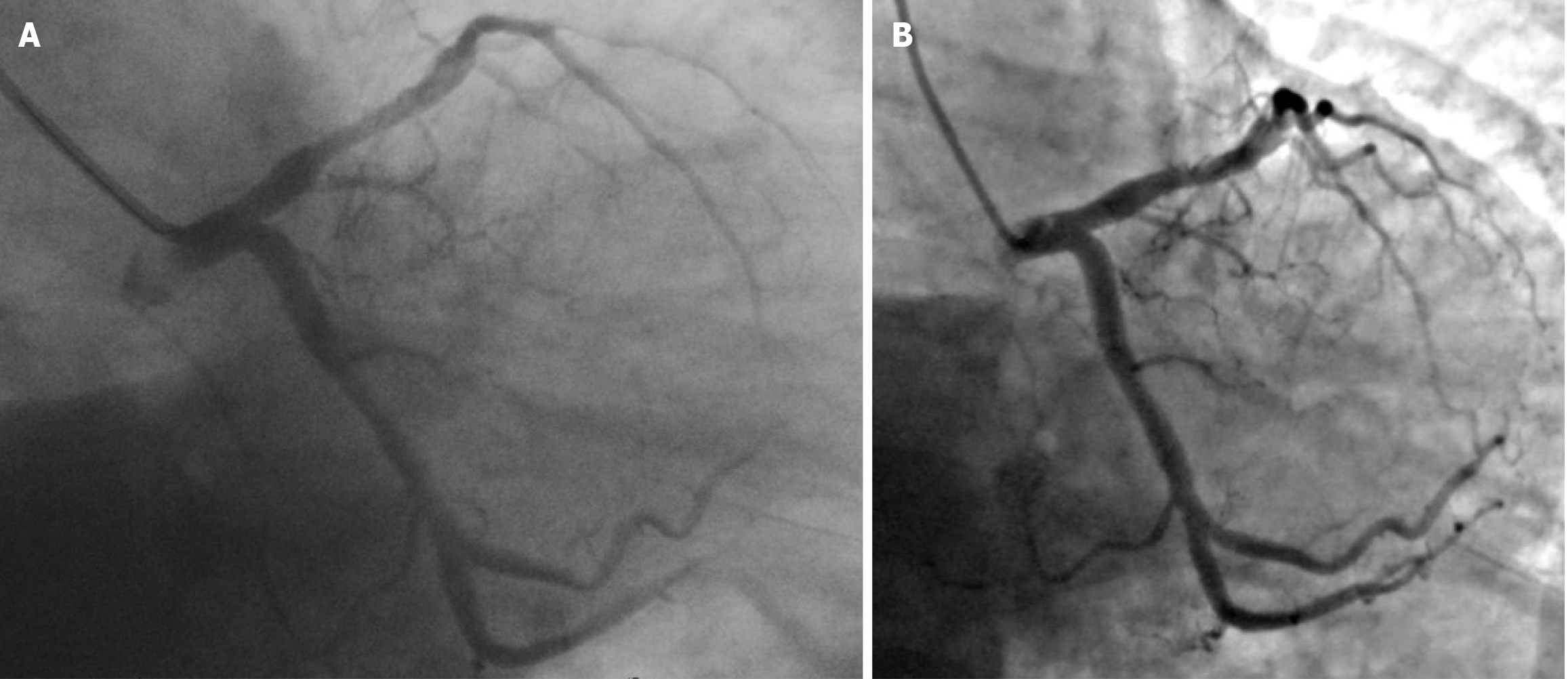
Figure 4 Comparison of the left coronary angiography images obtained in the same position.
A: On June 24, 2019, the residual stenosis in the proximal segment of the left anterior descending artery (LAD) after the primary percutaneous coronary intervention was approximately 40%, and the net lumen gain at the site of the most substantial stenosis was approximately 2.3 mm (RAO 30.0°, CAU 30.0°); B: On June 19, 2023, approximately 25% residual stenosis was observed in the proximal LAD, and the net lumen gain at the site of the most substantial stenosis was approximately 3.0 mm (RAO 30.4°, CAU 29.9°).
- Citation: She LQ, Gao DK, Hong L, Tian Y, Wang HZ, Huang S. Intracoronary thrombolysis combined with drug balloon angioplasty in a young ST-segment elevation myocardial infarction patient: A case report. World J Cardiol 2024; 16(9): 531-541
- URL: https://www.wjgnet.com/1949-8462/full/v16/i9/531.htm
- DOI: https://dx.doi.org/10.4330/wjc.v16.i9.531


